
The information outlined below on common conditions of the eye is provided as a guide only and it is not intended to be comprehensive.
Discussion with Mr Woodcock is important to answer any questions that you may have. For information about any additional conditions not featured within the site, please contact us for more information.
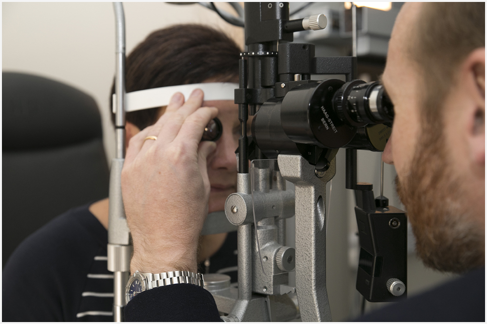
Common Conditions and Treatments of the Eye
During cataract surgery the natural lens of the eye, now cloudy with cataract, is removed. The natural lens is one of the two focusing elements of the eye, the other being the cornea. Replacing the cloudy natural lens with a transparent lens implant provides a perfect opportunity to improve the optical performance of the eye. Imperfections existing before the development of cataract may be treated using customised lens implants whose characteristics match those of the eye. Such imperfections include short sight, long sight, astigmatism and presbyopia (reading glasses dependence). These imperfections increase dependence on distance and reading glasses and reduce quality of vision, for example causing glare when driving at night.
< href=“conditions/cataract-surgery/“>Click here for more information.
Age-related macular degeneration (AMD) is a painless eye condition that causes you to lose central vision, usually in both eyes. Central vision is what you see when you focus straight ahead.
In AMD, this vision becomes increasingly blurred, which means:
reading becomes difficult
colours appear less vibrant
people's faces are difficult to recognise
This sight loss usually happens gradually over time, although it can sometimes be rapid. AMD doesn't affect your peripheral vision (side vision), which means it will not cause complete blindness.
WHEN TO SEEK MEDICAL ADVICE
Visit your GP or optometrist if your vision is getting gradually worse. If your vision suddenly gets worse, images are distorted or you notice blind spots in your field of vision, seek medical advice immediately and book an emergency appointment with an optometrist. If AMD is suspected, you'll be referred to an ophthalmologist (eye specialist) for tests and any necessary treatment.
WHY IT HAPPENS
Macular degeneration develops when the part of the eye responsible for central vision (the macula) is unable to function as effectively as it used to. There are two main types – dry AMD and wet AMD.
Dry AMD
Dry AMD develops when the cells of the macula become damaged by a build-up of deposits called drusen. It's the most common and least serious type of AMD, accounting for around 9 out of 10 cases.
Vision loss is gradual, occurring over many years. However, an estimated 1 in 10 people with dry AMD go on to develop wet AMD.
Wet AMD
Wet AMD – sometimes called neovascular AMD – develops when abnormal blood vessels form underneath the macula and damage its cells.
Wet AMD is more serious than dry AMD. Without treatment, vision can deteriorate within days.
WHO'S AFFECTED?
AMD currently affects more than 600,000 people in the UK and is the leading cause of vision loss. By 2020, it's predicted almost 700,000 people will have late-stage AMD in the UK.
For reasons that are unclear, AMD tends to be more common in women than men. It's also more common in white and Chinese people. The condition is most common in people over the age of 50. It's estimated 1 in every 10 people over 65 have some degree of AMD.
TREATING MACULAR DEGENERATION
There's currently no cure for either type of AMD. With dry AMD, treatment aims to help a person make the most of their remaining vision – for example, magnifying lenses can be used to make reading easier.
There's some evidence to suggest a diet rich in leafy green vegetables may slow the progression of dry AMD.
Wet AMD can be treated with anti-vascular endothelial growth factor (anti-VEGF) medication. This aims to stop your vision getting worse by preventing further blood vessels developing.
In some cases, laser surgery can also be used to destroy abnormal blood vessels.
The early diagnosis and treatment of wet AMD is essential for reducing the risk of severe vision loss.
REDUCING YOUR RISK
It's not always possible to prevent macular degeneration because it's not clear exactly what triggers the processes that cause the condition.
Your risk of developing AMD is closely linked to your age and whether you have a family history of the condition. However, you may be able to reduce your risk of developing AMD, or help prevent it getting worse, by:
stopping smoking if you smoke
eating a healthy, balanced diet that includes plenty of fruit and vegetables
moderating your consumption of alcohol
trying to achieve or maintain a healthy weight
wearing UV-absorbing glasses when outside for long periods
JUVENILE MACULAR DEGENERATION
In rare cases, macular degeneration can affect younger people. This is sometimes known as juvenile macular degeneration. It can be present at birth or develop later, but it's almost always caused by an inherited genetic disorder, such as:
Stargardt's disease – the most common cause of juvenile macular degeneration, this can start in childhood or early adulthood
Best's disease – also known as Best's vitelliform macular dystrophy
Sorsby's dystrophy – this often begins between the ages of 30 and 40
Vascular endothelial growth factor (VEGF), originally known as vascular permeability factor (VPF), is a signal protein produced by cells that stimulates vasculogenesis and angiogenesis. It is part of the system that restores the oxygen supply to tissues when blood circulation is inadequate. Serum concentration of VEGF is high in bronchial asthma and diabetes mellitus.[2] VEGF's normal function is to create new blood vessels during embryonic development, new blood vessels after injury, muscle following exercise, and new vessels (collateral circulation) to bypass blocked vessels.
When VEGF is overexpressed, it can contribute to disease. Solid cancers cannot grow beyond a limited size without an adequate blood supply; cancers that can express VEGF are able to grow and metastasize. Overexpression of VEGF can cause vascular disease in the retina of the eye and other parts of the body. Drugs such as bevacizumab and ranibizumab can inhibit VEGF and control or slow those diseases.
VEGF is a sub-family of growth factors, to be specific, the platelet-derived growth factor family of cystine-knot growth factors. They are important signaling proteins involved in both vasculogenesis (the de novo formation of the embryonic circulatory system) and angiogenesis (the growth of blood vessels from pre-existing vasculature).
Anti-VEGF therapies are important in the treatment of certain cancers and in age-related macular degeneration. They can involve monoclonal antibodies such as bevacizumab (Avastin), antibody derivatives such as ranibizumab (Lucentis), or orally-available small molecules that inhibit the tyrosine kinases stimulated by VEGF: lapatinib (Tykerb/Tyverb), sunitinib (Sutent), sorafenib (Nexavar), axitinib, and pazopanib. (Some of these therapies target VEGF receptors rather than the VEGFs.) THC and cannabidiol both inhibit VEGF and slow Glioma growth.[citation needed]
Both antibody-based compounds are commercialized. The first three orally available compounds are commercialized, as well. The latter two (axitinib and pazopanib) are in clinical trials.
Bergers and Hanahan concluded in 2008 that anti-VEGF drugs can show therapeutic efficacy in mouse models of cancer and in an increasing number of human cancers. But, "the benefits are at best transitory and are followed by a restoration of tumour growth and progression."
Later studies into the consequences of VEGF inhibitor use have shown that, although they can reduce the growth of primary tumours, VEGF inhibitors can concomitantly promote invasiveness and metastasis of tumours.
AZ2171 (cediranib), a multi-targeted tyrosine kinase inhibitor has been shown to have anti-edema effects by reducing the permeability and aiding in vascular normalization.
A 2014 Cochrane Systematic Review studied the effectiveness of ranibizumab and pegaptanib, on patients suffering from macular edema caused by central retinal vein occlusion. Participants on both treatment groups showed improvement in visual acuity measures and a reduction in macular edema symptoms over six months.
Vitreoretinal surgery refers to any operation to treat eye problems involving the retina, macula, and vitreous fluid. These include retinal detachment, macular hole, epiretinal membrane and complications related to diabetic retinopathy.
Click here for more information.
Diabetic retinopathy is a common complication of diabetes. It occurs when high blood sugar levels damage the cells at the back of the eye (known as the retina). If it isn't treated, it can cause blindness.
It's important for people with diabetes to control their blood sugar levels. Everyone with diabetes who is 12 years old or over should have their eyes examined once a year for signs of damage.
All people with diabetes are at risk of getting diabetic retinopathy, but good control of blood sugar levels, cholesterol and blood pressure minimises this risk.
How diabetes can damage the retina
The retina is the light-sensitive layer of cells at the back of the eye. It converts light into electrical signals.
The signals are sent to the brain through the optic nerve and the brain interprets them to produce the images that you see.
To work effectively, the retina needs a constant supply of blood, which it receives through a network of tiny blood vessels.
Over time, a continuously high blood sugar level can cause the blood vessels to narrow, bleed or leak. This damages the retina and stops it from working.
When the blood vessels in the central area of the retina (the macula) are affected, it's known as diabetic maculopathy.
Symptoms of diabetic retinopathy
During the initial stages, retinopathy does not cause any noticeable symptoms. You may not realise that your retina is damaged until the later stages, when your vision becomes affected. Vision loss will probably be permanent at this late stage, which is why diabetic eye screening is so important.
If you have diabetes and start to notice problems with your vision, contact your GP or diabetes care team immediately.
Screening for diabetic retinopathy
As severe retinopathy can cause sudden blindness, it needs to be identified and treated as soon as possible.
The NHS Diabetic Eye Screening Programme aims to reduce the risk of vision loss in people with diabetes. This is done by identifying retinopathy at an early stage and ensuring that treatment is given to reduce or prevent sight damage.
Everyone with diabetes who is 12 years old or over is invited for screening once a year.
The screening test involves examining the back of the eyes and taking photographs of the retina. Screening can detect diabetic retinopathy before you notice any changes to your vision.
Treating diabetic retinopathy
Treatment for retinopathy will depend on the stage the condition has reached.
For example, if retinopathy is identified in its early stages, you can prevent it from getting worse just by controlling your diabetes.
If you have more advanced retinopathy, you may need to have laser surgery or injection therapy to prevent further damage to your eyes.
Preventing diabetic retinopathy
To reduce your risk of developing retinopathy, it's important to control your blood sugar level, blood pressure and cholesterol level. Good control will prevent diabetic complications in almost everyone.
Other steps that you can take to help prevent retinopathy include:
attending your annual screening appointment
informing your GP if you notice any changes to your vision (do not wait until your next screening appointment)
taking your medication as prescribed
losing weight (if you're overweight) and eating a healthy, balanced diet
exercising regularly
giving up smoking
controlling your blood pressure and cholesterol levels
Mr Woodcock is fully qualified in this form of treatment for conditions at the back of the eye, which are treated medically using drugs, eye drops or lasers, and includes diabetic eye screening.
Conditions treated in this service include age-related macular degeneration (AMD), retinitis pigmentosa, diabetic retinopathy, retinal blood vessel blockages and inflammation at the back of the eye (uveitis).
Also known as primary care, Mr Woodcock and his team treat general eye problems and those that might need referral to one of our more specialist services.
Some eye problems don’t fit neatly into one of our other services, so our general clinics are an opportunity for our Mr Woodcock to make a full assessment and decide on the best treatment for you. You might also be assessed in this department if you have more than one eye condition.
Dry eye syndrome, or dry eye disease, is a common condition that occurs when the eyes do not make enough tears or the tears evaporate too quickly.
This leads to the eyes drying out and becoming inflamed (red and swollen) and irritated.
Dry eye syndrome is also known as keratoconjunctivitis sicca, or simply 'dry eyes'.
Symptoms of dry eye syndrome
The symptoms of dry eye syndrome usually affect both eyes and often include:
feelings of dryness, grittiness or soreness that get worse throughout the day
red eyes
eyelids that stick together when you wake up
temporarily blurred vision, which usually improves when you blink
See your GP or optometrist if you experience persistent symptoms of dry eye syndrome. They may examine you to check if the problem is caused by an underlying condition or may refer you to an eye specialist called an optometrist or ophthalmologist for further tests.
What causes dry eye syndrome?
Dry eye syndrome can occur when the complex tear production process is disrupted in some way. There are many different reasons why this can happen, although a single identifiable cause is not often found.
Common causes include:
being in a hot or windy climate
wearing contact lenses
certain underlying medical conditions, such as blepharitis (inflammation of the eyelids)
side effects of certain medications
hormonal changes, such as during the menopause (when a woman's periods stop)
Although the condition can affect people of any age, your chances of developing dry eye syndrome increase as you get older. It's estimated that up to one in every three people over the age of 65 experiences problems with dry eyes.
Dry eye syndrome is also more common in women than men.
How dry eye syndrome is treated
Dry eye syndrome is not usually a serious condition.
Treatments are available to help relieve the symptoms, which include eye drops to lubricate the eyes, medications to reduce any inflammation, and (if necessary) surgery to prevent tears from draining away easily.
If dry eye syndrome is caused by an underlying condition, treating this condition will usually help relieve the symptoms.
Caring for your eyes
As well as medical treatments, there are some things you can do yourself to help prevent dry eye syndrome or reduce the symptoms.
These include:
keeping your eyes and eyelids clean and protecting them from dusty, smoky, windy and dry environments
using your computer or laptop correctly to avoid eye strain
using a humidifier to moisten the air
eating a healthy diet that includes omega-3 fats
Further problems
Dry eye syndrome may be uncomfortable, but does not usually cause any serious problems. In rare cases, severe untreated dry eye syndrome can cause scarring of the eye's surface, leading to visual impairment.
Contact your GP or visit your nearest accident and emergency (A&E) department immediately if you have any of the following symptoms, as they could be a sign of a more serious condition:
extreme sensitivity to light (photophobia)
very red or painful eyes
a deterioration in your vision

A lazy eye, also known as amblyopia, is a childhood condition that occurs when the vision in an eye does not develop properly.
This usually means that the child can see less clearly out of one eye and relies more on the "good" eye.
An estimated 1 in 50 children will develop a lazy eye and children are usually diagnosed with the condition around the age of four.
How do I know if my child has a lazy eye?
A lazy eye does not usually cause symptoms. Younger children are often unaware that there is anything wrong with their vision and, if they are, they are usually unable to explain what is wrong. Older children may complain that they can't see as well through one eye.
In some cases you may notice that one eye looks different to the other. However, this is usually a sign of another condition that could lead to a lazy eye, such as a squint (when the eyes don't look in the same direction).
When to seek medical advice
Many cases of lazy eye are diagnosed during routine eye tests before parents realise that there is a problem. Children should have a vision test between the ages of four and five.
However, you should see your GP if you are concerned about your child's eyesight as they can refer your child for further testing by an eye specialist (opthalmologist).
What causes a lazy eye?
The eyes work like a camera. An image made up of light comes through the lens of each eye and is beamed onto a light sensitive layer of tissue called the retina.
The retina translates the image into nerve signals that are sent to the brain. The brain then combines the signals from each eye into a three-dimensional image.
A lazy eye occurs when the brain connections responsible for vision are not made properly. This can be because of:
a reduction in the amount of light entering the eye
a lack of focus in the eye
confusion between the eyes – where the two images are not the same (such as a squint)
Left untreated, this can lead to the central vision of the eye never reaching normal levels.
Treating a lazy eye
The majority of cases of lazy eye can be treated, usually in two stages.
The underlying problem is first corrected. This can be done using glasses to correct the focus of the eye, which often helps to correct a squint as well.
The child is then encouraged to use the affected eye again. This can be done with eye patches to cover the stronger eye, or using eye drops to temporarily impair the vision in the strong eye.
Treatment is often effective, but it's a gradual process, taking many months to work.
Watering eyes occur if too many tears are produced or if they cannot drain away properly.
The problem can affect anyone, but it's most common in young babies and people older than 60. It can cause blurred vision, sore eyelids and sticky eyes.
See your GP or optician if you have persistent watering eyes or any lumps or swelling around your eyes.
What causes watering eyes?
A problem with the glands
Glands in the eyelids (Meibomian glands) normally secrete an oily substance that slows the evaporation of tears between blinks.
When these glands don't function properly, known as Meibomian gland dysfunction (MGD), it can result in dry patches on your eyes. These become sore, and extra tears are produced as a reflex. This is the most likely cause of watering eyes.
Other causes
Other problems that can cause extra tears to be produced include:
the lower eyelid sagging away from the eye (ectropion) – this makes it difficult for tears to reach the drainage ducts
eyelids that roll inwards (entropion)
inflammation of the edges of the eyelids (blepharitis)
blocked or narrowed tear ducts
eye irritation (for example, from chemical fumes or grit)
an eye infection, such as conjunctivitis
an allergy
How are watering eyes investigated and treated?
Your GP may refer you to an optometrist (eye specialist) for an examination, if no obvious reason for your watering eye can be found.
Investigating the cause
If necessary, you may then be referred to an ophthalmologist (eye surgeon) for further investigation.
An ophthalmologist will look for blockages in your tear ducts, using local anaesthetic eye drops to help reduce any discomfort. This involves inserting a tiny probe into the narrow drainage channels on the inside of your eyelid to determine whether they are blocked. Fluid may also be injected into your tear duct, to see whether it comes out normally.
Another test involves placing a drop of special dye in each eye. If there is a significant amount left in your eyes after five minutes of normal blinking, your tear ducts may be blocked.
Scans of your tear ducts may sometimes be carried out. These can involve either injecting or placing special dyes into the tear ducts and then taking X-rays or other scans to help pinpoint the location of the blockage.
Treatment
If you're producing extra tears as a result of dry eye syndrome, you may be offered lubricating eye drops and advised to avoid activities that aggravate your symptoms.
Medication may be needed if the cause is an allergy or infection, and surgery may be needed if a tear duct is blocked.
If watering eyes aren't interfering with your life, you may choose not to have treatment.
The lacrimal system or apparatus is the anatomical system containing the ocular structures for tear production and drainage. Any disruption to either the production of tears or the drainage of tears will result in lacrimal disorders, which lead to acute or chronic discomfort of the eye.
DRY EYE
What is it?
drops for dry eye Dry eye is probably one of the most common problems seen in the ophthalmologist’s office. As we age, the protective tear film on the surface of the eye diminishes. This leaves the delicate tissues of the eye exposed to the drying effects of air, wind, dust and the sun. The eye can still make tears; in fact, many patients complain of wet eyes and tearing with this condition. This is due to the dryness producing a reflex tearing in an effort to keep the eye well lubricated.
What causes it?
In many people, the dryness is worse in the afternoon and evenings. Since we blink less frequently when we read, reading can also aggravate the symptoms of dry eyes.
Sometimes environmental factors play a role as well. Dry weather, either in hot or cold temperatures, robs the eye of needed lubricants. Cigarette smoke, fumes, dust and airborne particles are common irritants. In most patients, this condition is not associated with any systemic disease.
What are the symptoms?
Symptoms include burning, stinging, or a gritty sensation which may come and go depending on many factors. Itching, tearing, and light sensitivity may bother other patients. Occasionally long strings of mucus can be stretched from a dry eye. Actually, excessive watering of the eyes may indicate dry eyes, similar to the tearing which occurs with foreign material or lashes in the eye.
How it is treated?
Treatments helps in most patients. Because there is no cure, treatment must be ongoing. usually artificial tears, available over the counter, soothe the eyes and temporary relief. The disadvantage is that artificial tears only work for an hour or two, at best, and must be repeated at frequent intervals. Ointments last longer, but they blur vision and are most effective at night. Newer methods of treatment for seriously dry eyes are soft contact lenses in combination with artificial tears. Sometimes a slow-release medicine under the lower lid is helpful as well. Should symptoms persist, the drainage ducts can be temporarily or permanently closed, slowing the drainage of tears so they can soothe the dry eye.
Much research is being done on this subject because it is such a common problem. Time-release artificial tears seem to hold the most promise, but details of its use are still being worked out.
WET EYES
Blockage within the lacrimal drainage system can keep tears from draining into the nose, causing the tears to build up on the lower eyelid and spill over onto the face. The nasolacrimal duct, a bony canal carrying tears into the nose, is the most common site of obstruction. The main symptom is constant tearing from one or both eyes with tears running down the face. Because access into the nose is blocked, mucous builds up in the lacrimal sac making the patient prone to infection. Infection in the lacrimal sac can be serious as it can spread to the face, orbit and brain. This condition is treated initially with antibiotics followed by surgical correction of the obstructed duct.
BACTERIAL CONJUNCTIVITIS
The conjunctiva is the clear membrane that encircles and protects the eyeball. When you look at the white of the eye, you are really looking through the conjunctiva at the sclera, the tough, leathery outer coat of the eye. The conjunctiva has many small blood vessels running through it. The purpose of the conjunctiva is to lubricate and protect the eye and to allow it to move in its socket.
Conjunctivitis is an inflammation of the lining of the eye. It can be caused by bacteria (as in “pink eye”), viruses, chemicals, allergies, and more. It is sometimes difficult to tell exactly which is the real cause.
Bacterial conjunctivitis is characterized by swelling of the lid, a yellowish discharge, sometimes a scratchy feeling in the eye, and itching and mattering of the lids, especially in the mornings upon awakening. The conjunctiva is red and sometimes thickened. Often both eyes are involved. The bacteria most commonly at fault are the staphylococcus, the streptococcus, and H. influenza. This disease is very contagious, and sometimes entire families are infected. Laboratory cultures are not typically used to make the diagnosis since this is expensive and time consuming. Most infections are over by the time the results of the lab tests come back.
Treatment is curative. Usually antibiotic drops and compresses ease the discomfort and clear up the infection in just a few days.
Occasionally, the infection does not respond well to the drops. In those rare cases, a second visit to the office should be made and other measures undertaken. In severe infections, oral antibiotics are necessary. Covering the eye is not a good idea because that incubates the germs. If left untreated, conjunctivitis can create serious complications, such as infections in the cornea, lids, and tear ducts.
Prevention is important for avoiding the disease and stopping its spread. Careful washing of the hands, the use of clean handkerchiefs, and avoidance of contagious individuals are all helpful. Little children frequently get conjunctivitis because of their lack of understanding about hygiene and a resulting contact with germs.
CHALAZION
Along the upper and lower lids are located a number of glands that manufacture part of the tear film that protects and lubricates the eyeball. If one of these glands becomes blocked, a small lump forms. This is called a chalazion (chalazia, plural).
Chalazia may vary in size from small, almost invisible lumps to rather large masses as big as a little fingernail. Sometimes tender in their early stages, they are later painless and frequently will form a firm swelling in the lid. This lump can distort the eyeball, causing blurred vision if left untreated.
Chalazia are not caused by infection; however, they may become a site for infection once they have become established. Their exact cause remains unknown. Several conditions are associated with chalazia: seborrhea, chronic lid inflammation, dry eyes, and acne.
Most chalazia will disappear in a few weeks without any special therapy. To help them go away, frequent hot packs throughout the day and drops are helpful, especially in the early stages. In some cases, oral medicines can help prevent recurrences. If a chalzaion persists, a simple in-office surgical procedure can be performed to remove it. The chalazion is drained from the inside of the lid after a small injection of a local anesthetic. There is no visible scar and healing is rapid and painless. Often the eye is patched overnight to ensure proper healing.
Patient Information Leaflets